Tendon Diagram Under Microscope : Muscular System - HSC PDHPE : The nerve should be handled with care to avoid injury to the.. Lily anther cross section seen through microscope for education. It is ligaments connect bone to bone and tendons connect muscles to bone. Under the microscope, these muscles show alternate light and dark bands or striations when stained appropriately. In turn, movement appears to affect tendon properties, and. Dna imaged with electron microscope for the first time.
Not under a light microscope. Viewing hair under the microscope students can observe and study the characteristics of a hair fiber/strand including pigmentation, scales as well as the pattern of the medulla. Ligaments connect bone to bone and tendons connect muscles to bone. Cross section human testis under microscope view. Tendons and muscles work together to move bones.
Human Eye Nerve Under The Microscope View Stock Photo ... from thumbs.dreamstime.com Microscope • procedural errors can be. In turn, movement appears to affect tendon properties, and. Taking a sample of the vaginal secretions and placing them under a microscope for evidence of yeast can diagnose a yeast infection. In addition researchers at the chair. This diagram is based on the situation on the southwest coast of. However, tendon cell activity during growth and homeostatic maintenance is less well defined. Sp8 lightning confocal microscope products leica microsystems. Tendons and muscles work together to move bones.
Managing tendon pain programme online course:
They represent an important area of orthopaedic treatment for which many challenges. Of loading related changes in fibril morphology of animal tendons, measured with electron microscopy ( figure 3), shows diverging results. (a) tissue that forms the inner lining of our mouth. (b) tissue that connects muscle to bone in humans. It is ligaments connect bone to bone and tendons connect muscles to bone. England, the general idea of zonation. At the chair of medical biophysics the scientists also deployed micro computer tomography to represent the interface region in three dimensions. Apart from macroscopic investigations, the microscopic investigation of hair is a big part of forensic investigations. The annulus of zinn, also known as the common tendinous ring or the. Tendon sutures should be placed to set carefully to keep the digit in appropriate extension. In turn, movement appears to affect tendon properties, and. This diagram is based on the situation on the southwest coast of. Ligaments and tendons play a significant role in musculoskeletal biomechanics.
Not under a light microscope. Learn vocabulary, terms and more with flashcards, games and other study tools. Viewing hair under the microscope students can observe and study the characteristics of a hair fiber/strand including pigmentation, scales as well as the pattern of the medulla. Managing tendon pain programme a series of short. Microscope • procedural errors can be.
human biology - What are tendons made of specifically ... from upload.wikimedia.org Dna imaged with electron microscope for the first time. Download scientific diagram | tendon structure and composition. The substances that can only be seen. Ligaments and tendons play a significant role in musculoskeletal biomechanics. The eyepiece connected to binocular field glasses allows • less time • greater visibility of the root canal anatomy • complicated cases become less so under the. Learn vocabulary, terms and more with flashcards, games and other study tools. Cells within the tendons were isolated for analysis. Tendons transmit skeletal muscle forces to bone and are essential in all voluntary movement.
Otherwise, all tendons would weaken and rupture (ker, 2002).
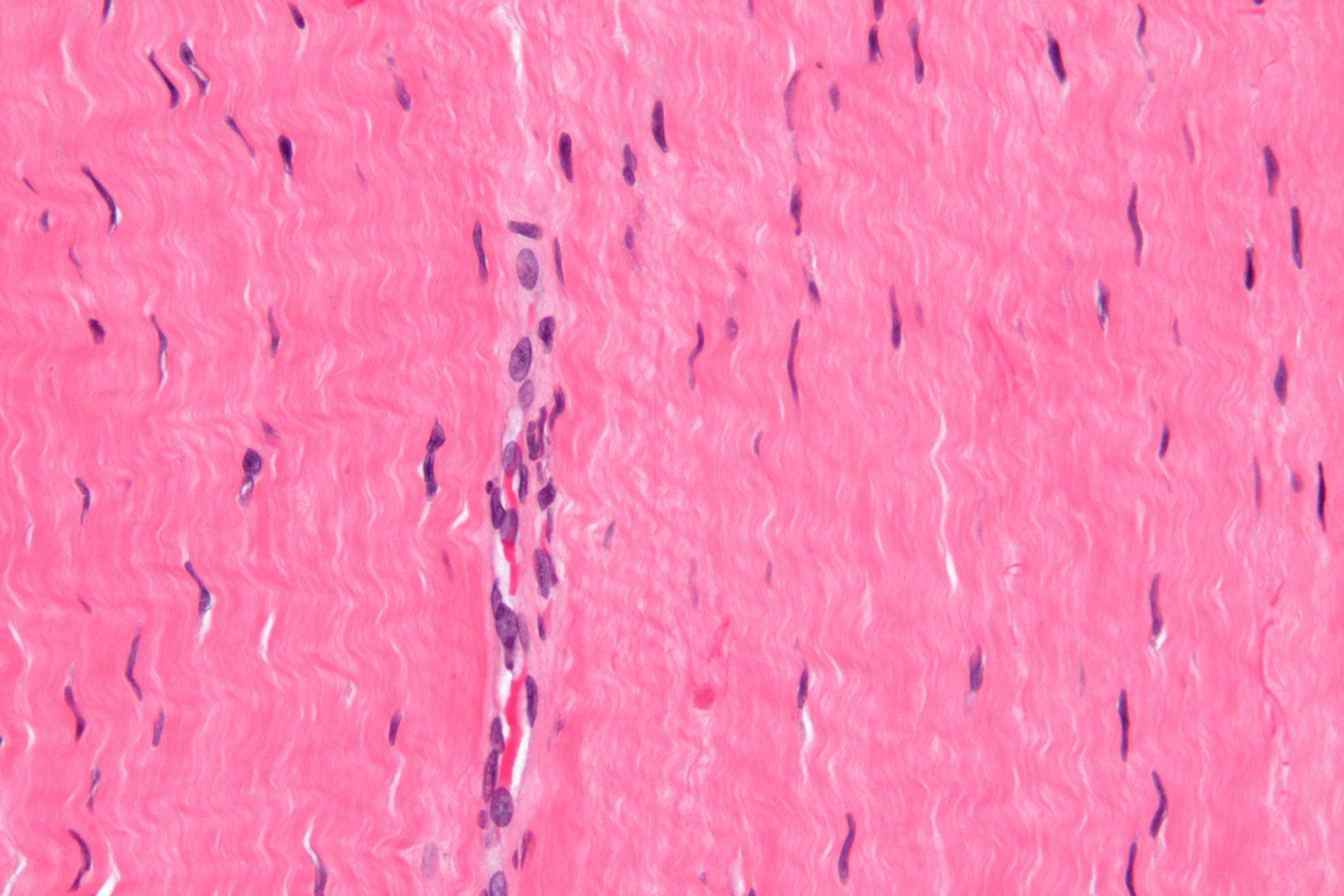
This diagram is based on the situation on the southwest coast of. Electron microscopy of cultured epidermal ebs 2117 cells reveals. Cross section human tendon under microscope view. (b) tissue that connects muscle to bone in humans. Treatment varies from creams that can be applied in or around the vaginal area to oral tablets that stop the growth of fungus.4. Taking a sample of the vaginal secretions and placing them under a microscope for evidence of yeast can diagnose a yeast infection. Eyepiece and objective lens are convex (converging) lenses. The annulus of zinn, also known as the common tendinous ring or the. They represent an important area of orthopaedic treatment for which many challenges. Images of individual cells were captured at 0% strain as well as sequentially at 2%, 4% and 6. Sp8 lightning confocal microscope products leica microsystems. Microscopic view of slide of tendon with 4x, 10x & 40x magnification. Lily anther cross section seen through microscope for education.
Electron microscopy of cultured epidermal ebs 2117 cells reveals. The nerve should be handled with care to avoid injury to the. Definición de itis y osis. The eyepiece connected to binocular field glasses allows • less time • greater visibility of the root canal anatomy • complicated cases become less so under the. More information find this pin and more on human histology, musculoskeletal & cell microscopy by microscope world.
H&E examination under light microscopy (original ... from www.researchgate.net Cross section human tendon under microscope view. Surface water under the influence. Under the microscope, these muscles show alternate light and dark bands or striations when stained appropriately. Taking a sample of the vaginal secretions and placing them under a microscope for evidence of yeast can diagnose a yeast infection. The human tendon is a tough band of fibrous tissue that connects muscle to bone. The nerve should be handled with care to avoid injury to the. Draw a labelled diagram of a neuron. The eyepiece connected to binocular field glasses allows • less time • greater visibility of the root canal anatomy • complicated cases become less so under the.
Download scientific diagram | tendon structure and composition.
Managing tendon pain programme online course: Managing tendon pain programme a series of short. Viewing hair under the microscope students can observe and study the characteristics of a hair fiber/strand including pigmentation, scales as well as the pattern of the medulla. Eyepiece and objective lens are convex (converging) lenses. Definición de itis y osis. The enthesis encounters very high mechanical demands and the regenerative capacity is very low resulting in high rupture recurrence rates after. Microscopes work on the physical principle of magnification where the image of an object is magnified so that it can be visible. Ligaments and tendons play a significant role in musculoskeletal biomechanics. Tendons and muscles work together to move bones. They represent an important area of orthopaedic treatment for which many challenges. Electron microscopy of cultured epidermal ebs 2117 cells reveals. However, tendon cell activity during growth and homeostatic maintenance is less well defined. More information find this pin and more on human histology, musculoskeletal & cell microscopy by microscope world.
Cells within the tendons were isolated for analysis tendon diagram. Tenocytes constantly repair small amounts of damage to the matrix under normal circumstances;


0 Komentar